Functional Magnetic Resonance Imaging (fMRI)
A Brief History

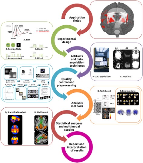
Functional Magnetic Resonance Imaging (fMRI) is a non-invasive brain imaging technique that measures changes in blood flow in response to neural activity. The history of fMRI can be traced back to the 1980s when researchers first began to explore the use of MRI to investigate brain function. In 1990, a team led by Seiji Ogawa discovered that the signal from MRI scans could be affected by changes in blood flow, and that this signal change could be used to infer neural activity. This discovery led to the development of fMRI, which quickly became a popular tool for studying brain function. The first fMRI experiments were performed in the early 1990s, and by the mid-1990s, fMRI had become widely used in cognitive neuroscience. Since then, fMRI technology has continued to improve, with advances in hardware and software allowing for higher resolution and more detailed images of the brain. Today, fMRI is a standard tool used in neuroscience research, and has also found clinical applications in the diagnosis and treatment of neurological and psychiatric disorders.
Blood Oxygen Level Dependent (BOLD) Signals

BOLD (Blood Oxygenation Level Dependent) signals are changes in the magnetic properties of hemoglobin in the blood that occur when blood oxygen levels change in response to neural activity. When neurons become active, they require more oxygen, which leads to an increase in blood flow to the activated brain region. The increased blood flow brings more oxygenated hemoglobin to the area, causing a local increase in magnetic susceptibility that can be detected by fMRI scanners. This signal change is what fMRI researchers refer to as the BOLD signal. By measuring the BOLD signal, fMRI can provide information about which areas of the brain are active during a particular task or state, and how these areas are connected to one another.
There are several factors that can affect the BOLD signal in fMRI studies.
Analysis of fMRI

