Magnetoencephalography (MEG)
A Brief History
Magnetoencephalography (MEG) is a technique that measures the magnetic fields generated by the electrical activity of the brain. The history of MEG can be traced back to the early 20th century when the first magnetic field measurements were made using coils of wire to detect the small magnetic fields generated by the human heart.
In the 1960s, David Cohen and others began to develop MEG as a method for studying brain function. They discovered that the magnetic fields generated by the brain were much smaller than those generated by the heart, so they needed to develop highly sensitive detectors. In the 1970s and 1980s, several groups around the world developed MEG systems that were capable of measuring the magnetic fields generated by the brain. These systems used superconducting quantum interference devices (SQUIDs) to detect the magnetic fields.
Since then, MEG has been used to study a wide range of brain functions, including sensory processing, language, memory, and motor control. It has also been used to study neurological and psychiatric disorders such as epilepsy, schizophrenia, and autism. In recent years, advances in MEG technology have led to improvements in spatial and temporal resolution, making it an increasingly important tool for studying the brain. In particular, MEG has a unique strength as a functional brain imaging tool with high spatial-resolution comparable to functional MRIs.
Origins of MEG
MEG and EEG signals are both measures of the electrical activity in the brain, but they differ in the way that they are measured. EEG measures the electrical activity of the brain using electrodes placed on the scalp, while MEG measures the magnetic fields generated by this activity using a SQUID that is highly sensitive sensor. The origins of both MEG and EEG signals lie in the net effect of ionic currents flowing in the dendrites of neurons during synaptic transmission. Any electrical current generates a magnetic field, and conversely, any magnetic field generates an electrical current. Therefore, as neurons in the brain communicate with each other, they generate both electrical and magnetic fields.
While EEG measures the electrical activity of the brain, MEG measures the magnetic fields generated by this activity. The magnetic fields are generated by the same ionic currents that generate the electrical fields measured by EEG, but they are much weaker in magnitude. This is because the magnetic fields are generated by the net effect of ionic currents, while the electrical fields are generated by individual ionic currents. Overall, while MEG and EEG measure different aspects of the same underlying electrical activity in the brain, they provide complementary information that can be used together to gain a more complete understanding of brain function.
SQUID Sensors of MEG System

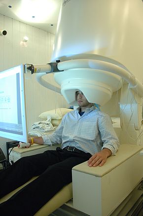
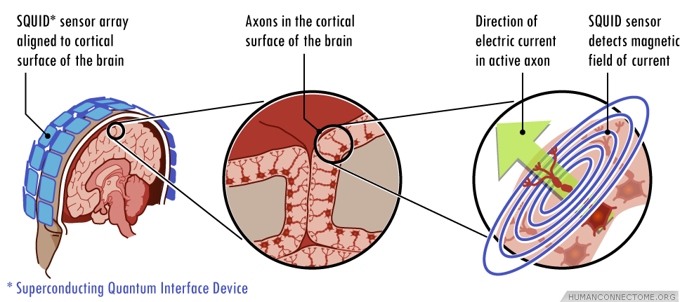
MEG sensors are based on the principle of superconductivity, which means they require very low temperatures to operate. The sensors are usually made of a thin layer of a superconducting material, such as niobium or lead, that is cooled to a temperature close to absolute zero (-273°C) using liquid helium. MEG helmet contains an array of liquid-helium-cooled low critical-temperature (low-Tc) SQUIDs that surround the head and sample the magnetic field generated from neural currents. Neuromagnetic field decays as a function of distance between the SQUID sensor and the neural source. Therefore, the closer the sensors are to the head, the better the SNR and spatial resolution for MEG.
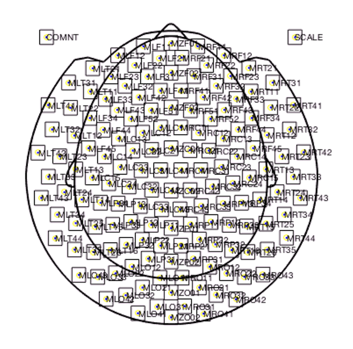
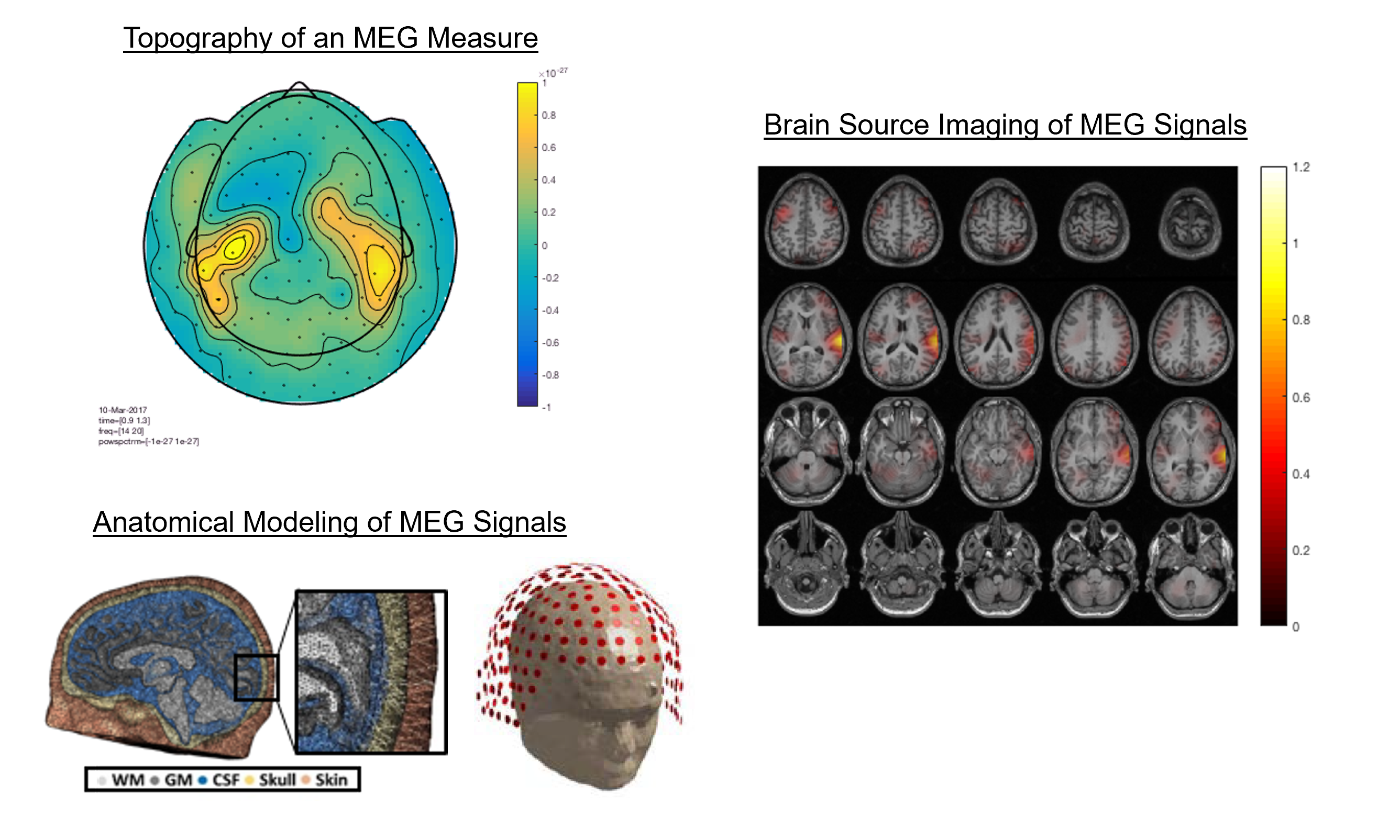
The sensors work by detecting changes in the magnetic field as the brain's electrical activity flows through neurons. The magnetic fields produced by these electrical currents are very weak and require highly sensitive sensors to detect them. There are two main types of MEG sensors: magnetometers and gradiometers. Magnetometers measure the strength of the magnetic field, while gradiometers measure the changes in the magnetic field over a distance. Gradiometers are more sensitive than magnetometers and can detect smaller magnetic fields, which makes them more commonly used in MEG systems. Overall, MEG sensors are a powerful tool for studying brain function and are used in a variety of research applications, including neuroscience, psychology, and clinical medicine. MEG is often used to measuring the brain activities and localize the sources in the brain. For accurate brain source localization, MEG systems tends to have large number of sensors (>128) in the helmet.
Domains of MEG Data Analysis
Although MEG and EEG represent different physical measures of neural activities (i.e., magnetic fields vs. electrical voltage), their signals share largely common features. They are direct and non-invasive measure of neural activities recorded at a high sampling rate (~1000 Hz). Therefore, all the EEG analysis techniques can be applied to MEG data in the time, frequency, time-frequency, and spatial domains.
Time domain

Frequency domain
Time-Frequency domain

Spatial domain